Researchers use pig ultrasounds at county fairs for education purposes
August 2, 2012
The word “ultrasound” is commonly used when referring to a woman’s pregnancy. Many people do not think of it as being used with animals. However, that is precisely what Iowa State has been using ultrasounds for since the 1990s.
“Real-time ultrasound has been around since the 1970s,” said Chad Yoder, graduate student in animal science. Yoder has been involved with the program since June 2010. “The equipment is similar to what is used in hospitals for pregnancy ultrasounds, and we actually use hospital equipment (though out of date) for ultrasounding. Iowa State began the county fair ultrasound program in the early 1990s.”
Yoder works with Colin Johnson, extension program specialist with the Iowa Pork Industry Center, to perform the ultrasounds on swine being shown at many of the county fairs in Iowa.
Ultrasounds are often performed at county fairs on swine, as well as sheep and cattle in some organizations.
Thus, it makes for a busy summer for the team.
Yoder also has to balance ultrasounds at the county fairs along with research projects for his graduate program.
“It is quite hectic to try and fit in research projects for graduate school and all of the county fairs, which leads to many long days, but fortunately, Iowa State does not ultrasound all 99 counties,” Yoder said. “Not all counties request this service, and there are several companies in Iowa that provide this service as well. Also, most county fairs occur during the same three-week period in July, which would make it impossible for us to do them all since Iowa State currently has three certified technicians. Iowa State was scheduled to ultrasound at 36 county fairs this year as well as the state fair.”
Yoder said the main use is as a tool for genetic improvement within the swine industry.
“Ultrasound was developed as a tool to measure traits on a live animal that could previously only be measured on the carcass post-mortem,” Yoder said. “For example, in pigs we can measure the amount of back fat the pig has, a measure of its leanness; how big its loin muscle area is, a measure of muscle or lean tissue in the pig; or intramuscular fat, the fat within the muscle that provides the flavor, also known as marbling, which is also used as a measure of pork quality. Now we can measure them on the animal and select the animals to be parents that excel in these traits so their progeny have higher genetic potential for these traits.”
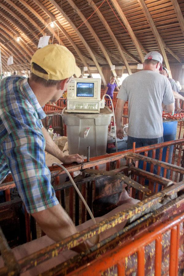
When going to the county fairs, it typically takes a few hours for Yoder and Johnson to perform the ultrasounds on all the swine.
“Each pig only takes about a minute,” Johnson said. “[But] most fairs have anywhere from 20–150 pigs for ultrasounding.”
Fair exhibitors are often in charge of taking their pigs and corralling them into the chutes for ultrasounding.
“First, we have the exhibitors get their pigs up here, and one pig at a time is placed in the chute,” Johnson said. “Then we pour a little bit of vegetable oil on the pig’s back, and then we place this transducer on the pig’s back. We want to try to measure in the space between the pig’s 10th and 11th ribs.”
Yoder said it can be difficult to figure out where those ribs are, but it’s made much easier once the ultrasound transducer, or the “probe” used to see the muscle and fat, is put on the pig’s back. Once this is placed, it can be adjusted slightly in order to find the space between the 10th and 11th ribs.
“Once the transducer is on the pig it can be adjusted slightly to find the space between the ribs. The reason we chose that location is because there are landmarks inside the pig’s muscle that tell us we are in that exact location and not too far forward or too far backward.”
Yoder said they try to find the same place on every pig so the comparisons are accurate.
“Finding it is not as difficult as it may seem,” Yoder said. “If I were to be too far forward — at the ninth rib for example — there would be an extra muscle in the picture — the spinalis muscle — and the shape would be different than it should look. If I were too far back — at the eleventh/twelfth rib — then the shape would be very distorted, so we know what to look for every time.”
Once the ideal image is received, a button is clicked to freeze it. Then the pig is let out of the chute and returned to its pen. Next the image is traced to determine loin muscle area and fat depth. The machine calculates the area and depth.
“The main reason we go to county fairs is to educate exhibitors,” Yoder said.
“We want the exhibitors to understand what we are doing, why we are doing it and what the value is to the commercial swine industry. These are measures of interest to the entire swine industry, just at the fair level there are awards for it. The point of these measures at the fair is to relate what the exhibitor has done with their animal to what is occurring in the commercial swine industry and mainly as an educational tool to prepare the exhibitors that may want to eventually pursue a career in the swine industry.”